
A normal semen analysis followed by total fertilization failure. Textbook ICSI on a mature oocyte, and no pronuclei the next morning. A Day 3 embryo that graded well and then stopped. Two patients, identical protocols, very different outcomes.
None of these are technique problems. That is what makes them frustrating, and it is also what makes them explainable. In every one of those cases, the answer sits below what the microscope shows us, in the cell biology and molecular biology that we tend to treat as coursework we already finished.
When I was a student, there was a question I kept returning to, and I suspect many people entering embryology ask it quietly without saying it aloud: why are we studying organelles again?
I had already covered membranes, DNA, mitochondria and metabolism during my Biotechnology degree. Entering Clinical Embryology, it felt for a while as though I was repeating material I had finished. Then I began working with real gametes and real embryos, and the separate chapters collapsed into one continuous story.
In the IVF laboratory, cell biology stops being a subject. It becomes the explanation for everything on the bench in front of you. Why the incubator must hold its temperature. Why a semen analysis can look acceptable and fertilization can still fail. Why an embryo that graded beautifully on Day 3 can arrest on Day 4. Why a woman of thirty-eight and a woman of twenty-eight can receive identical care and face very different odds.
This is what I want to write about. Not as a textbook summary, but as the reasoning I use every day.
1. We were all once a single cell
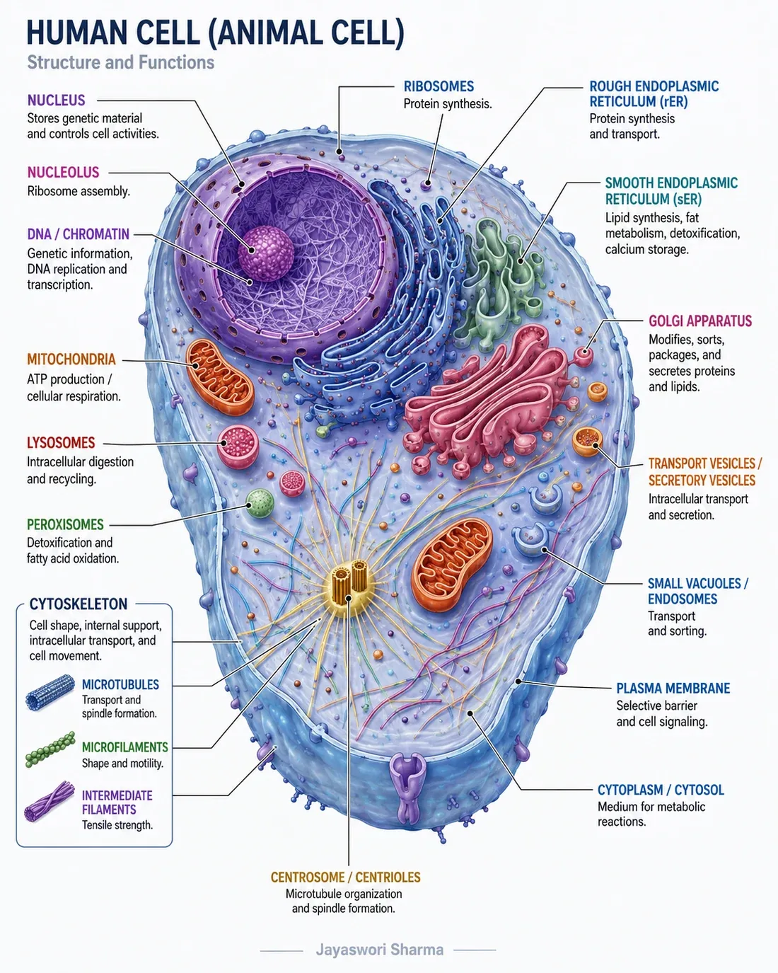
Meiosis creates haploid gametes. Fertilization restores the diploid genome. Mitosis then turns one zygote into a multicellular embryo. Three processes, and between them they account for the entire beginning of a human being.
What strikes me is how unequal the two contributions are. The oocyte provides almost all of the cytoplasm, along with maternal RNA, proteins and mitochondria. The sperm contributes paternal DNA and, in humans, centrosomal components that help organize the first divisions. One cell brings the instructions. The other brings the instructions and the entire factory.
Inside that factory, every structure has a job that matters clinically. The membrane controls ions, nutrients, water and pH. The nucleus manages genetic information. Mitochondria produce ATP. The endoplasmic reticulum, ribosomes and Golgi build and process molecules. The cytoskeleton moves chromosomes and physically divides the cell.
Even ovarian stimulation, which we tend to think of as a clinical protocol rather than cell biology, is cell signalling. FSH and LH bind their receptors and activate intracellular pathways that change steroid production and gene expression. When we adjust a stimulation protocol, we are adjusting molecular events inside granulosa cells.
Key idea: A small change outside the cell can become a major change inside it. This is exactly why temperature, pH, osmolality and air quality matter so much in an IVF laboratory.
2. A motile sperm is not yet a fertilizing sperm
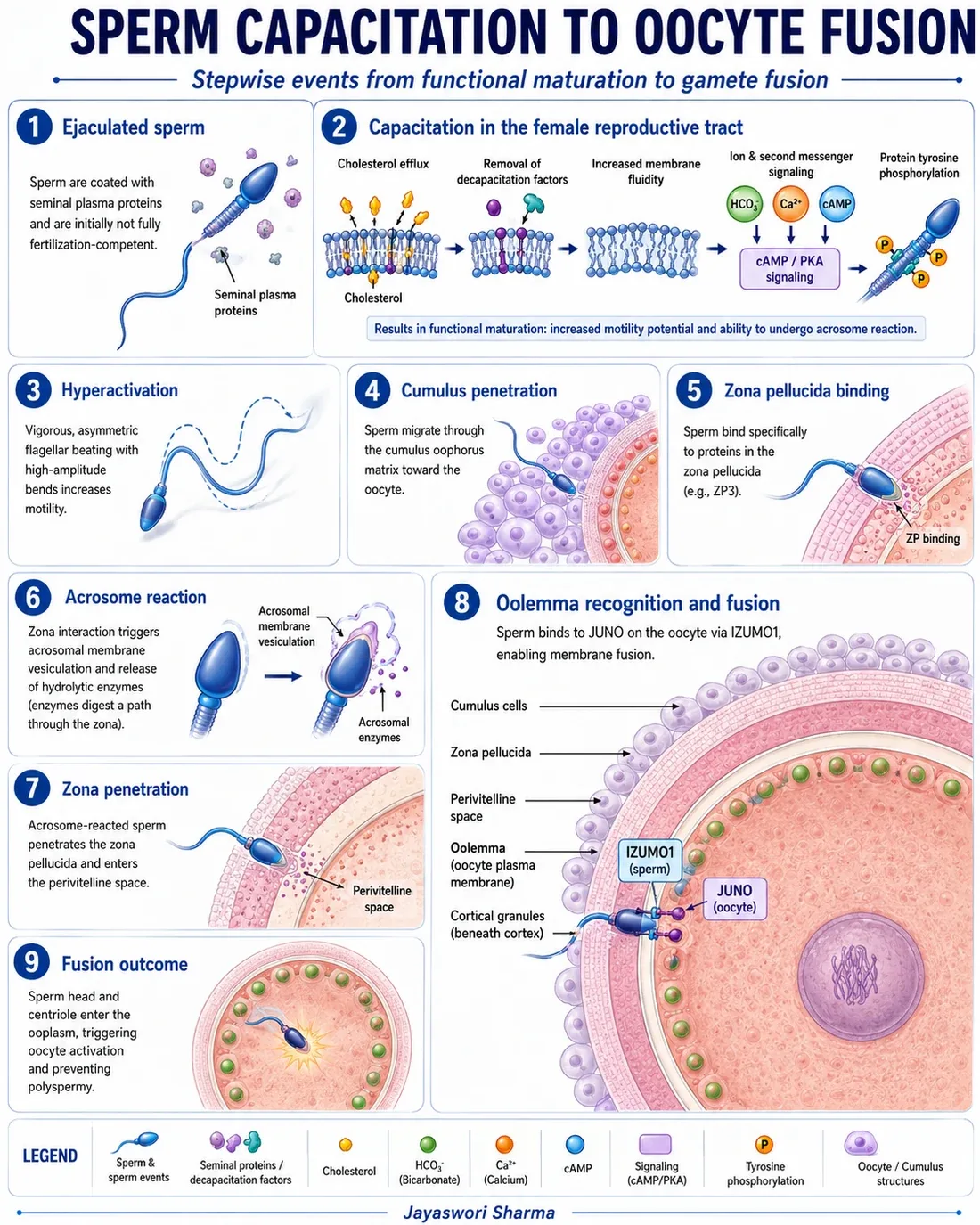
This is the point I wish I had understood earlier in my career, because it changes how you read a report.
After ejaculation, sperm is not yet fully prepared to fertilize. Capacitation has to happen first, and capacitation is a molecular remodelling: changes in membrane cholesterol, changes in ion movement, changes in protein phosphorylation.
Those changes are what produce hyperactivated motility, prepare the acrosome reaction, support penetration of the cumulus and the zona pellucida, and finally make membrane fusion possible. At the oolemma, IZUMO1 on the sperm and JUNO on the oocyte are important recognition partners within a larger fusion system.
There is also an asymmetry in gamete production worth pausing on. One primary spermatocyte can form four sperm. One primary oocyte usually forms one large oocyte and small polar bodies. The female pathway deliberately sacrifices cell number to preserve the cytoplasm that early development will depend on. Nothing about that is accidental.
The clinical consequence is uncomfortable but important. A sperm sample may look entirely acceptable in count and motility and still fail during capacitation, during the acrosome reaction, during zona interaction, or at fusion. Fertilization failure with a normal semen report is not a contradiction. It is a reminder of what the report does not measure.
Key idea: A semen analysis describes visible performance. Cell biology explains whether the sperm is functionally ready to fertilize.
3. ICSI places the sperm inside, but activation is what starts development
One injection does not automatically equal one activated oocyte. I have seen technically flawless ICSI followed by fertilization failure, and the explanation is not in the micromanipulator.
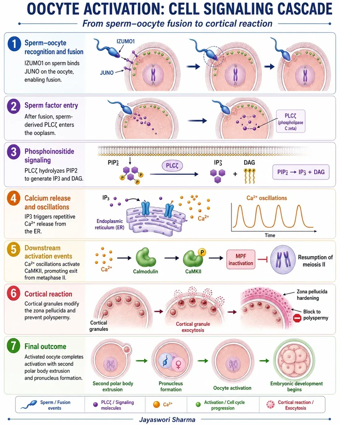
After sperm and oocyte fusion, or after sperm entry during ICSI, sperm-derived PLC zeta initiates IP3 signalling. IP3 releases calcium from the endoplasmic reticulum in repeated oscillations, not as a single spike. Calmodulin and CaMKII then translate that calcium pattern into a cell-cycle response.
Only then does the oocyte complete meiosis II, extrude the second polar body, release its cortical granules, form pronuclei and prepare for the first cleavage. The cortical reaction changes the zona pellucida and helps prevent additional sperm from entering.
So fertilization can fail after correct ICSI for two distinct reasons: the sperm activation signal was inadequate, or the oocyte could not respond properly. Those are different problems with different implications for the next cycle, and we can only separate them if we are thinking at this level.
Key idea: ICSI is a laboratory procedure. Oocyte activation is a coordinated molecular event. They are not the same thing.
4. The embryo must move from maternal control to its own program
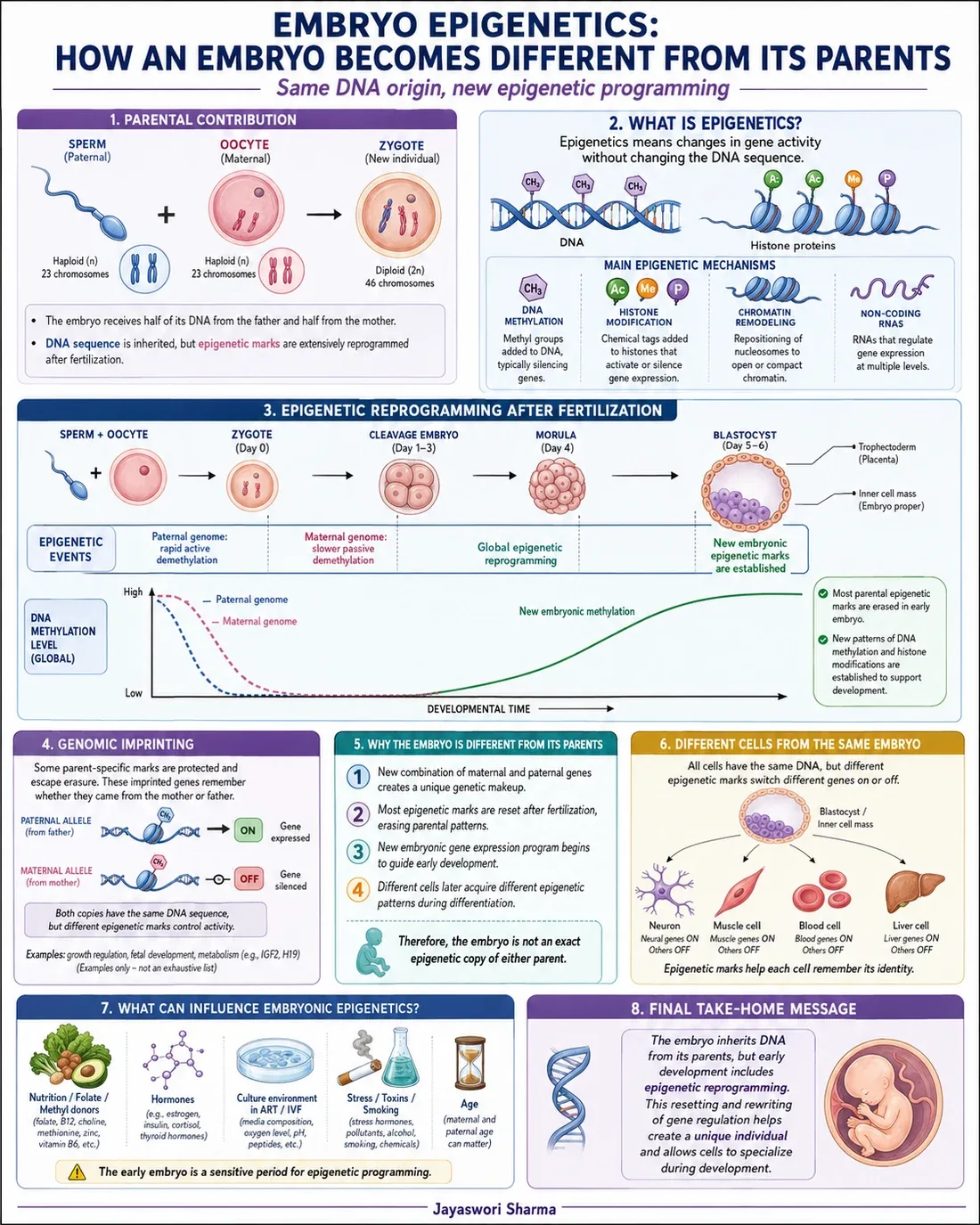
The early embryo does not become independent immediately, and I think this is one of the most underappreciated facts in routine embryo assessment.
The first cleavages depend heavily on maternal messenger RNA, proteins, ribosomes, organelles and metabolites that were stored inside the oocyte long before fertilization. The embryo is running on supplies, not on its own production.
Human embryonic transcription begins gradually, while major embryonic genome activation becomes prominent during the four-cell to eight-cell period. Around the same time, epigenetic reprogramming reorganizes DNA methylation, histone marks, chromatin accessibility and non-coding RNA.
The DNA sequence is inherited from the parents, but the way that DNA is used is reorganized. This is what allows cells carrying an identical genome to become inner cell mass, trophectoderm, placenta and fetal tissues. Differentiation is not a change in the code. It is a change in which parts of the code are read.
Clinically, this is why an embryo can appear morphologically acceptable and still arrest. If maternal RNA clearance, genome activation or epigenetic regulation is disturbed, the appearance under the microscope may lag behind the failure that has already occurred.
Key idea: The embryo does not only divide. It must learn to control its own genome.
5. Metabolism is not only fuel, it is also information
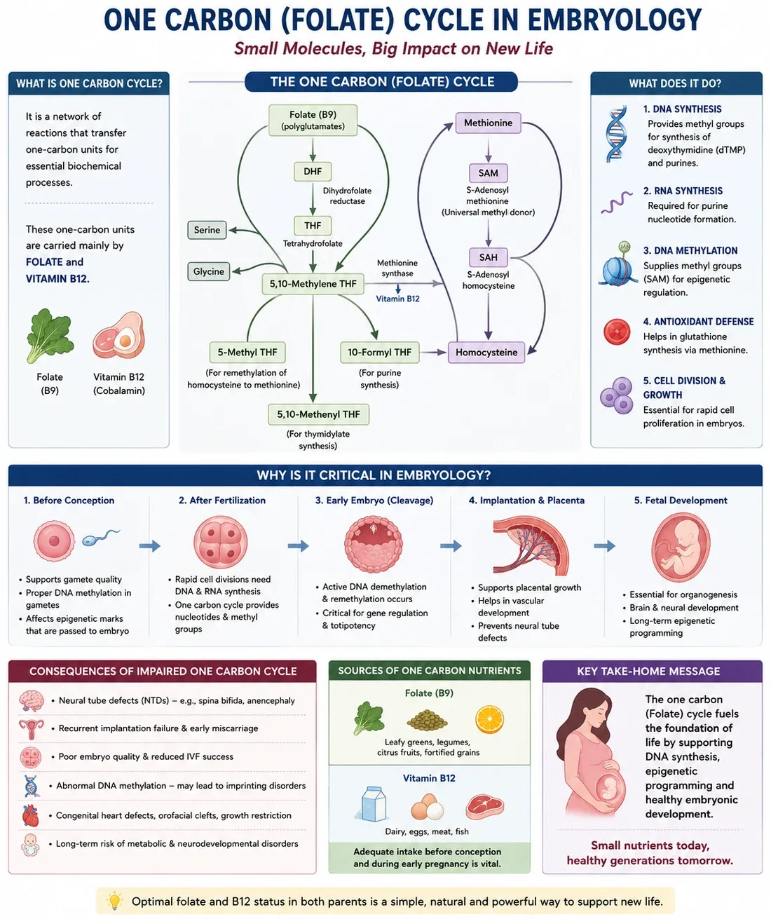
A nutrient can become a nucleotide, an antioxidant substrate, or an epigenetic mark. Once I understood that sentence, I stopped thinking of metabolism as a separate topic.
The folate cycle transfers one-carbon units. The connected methionine cycle produces S-adenosylmethionine, or SAM, the major cellular methyl donor. SAM then supplies methyl groups for DNA, histones, RNA and proteins.
Folate metabolism also supports purine and thymidylate synthesis, which are required to copy DNA during cell division. So the same pathway feeds both the writing of epigenetic marks and the physical duplication of the genome.
Taken together, the methionine and folate pathways connect nutrition with cell proliferation, antioxidant defence, genomic imprinting and embryonic gene regulation. This is why metabolism cannot be separated from molecular embryology. The embryo uses small molecules both as fuel and as instructions for regulation.
The clinical translation needs care, and I want to be precise here rather than enthusiastic. Balanced folate, vitamin B12 and methionine metabolism matters. Deficiency can be harmful. But unnecessary excess is not automatically beneficial, and supplementation should not be treated as a competition.
Key idea: One-carbon metabolism is the bridge between nutrients, DNA synthesis and epigenetic programming.
6. Reactive oxygen species can support fertilization or damage it
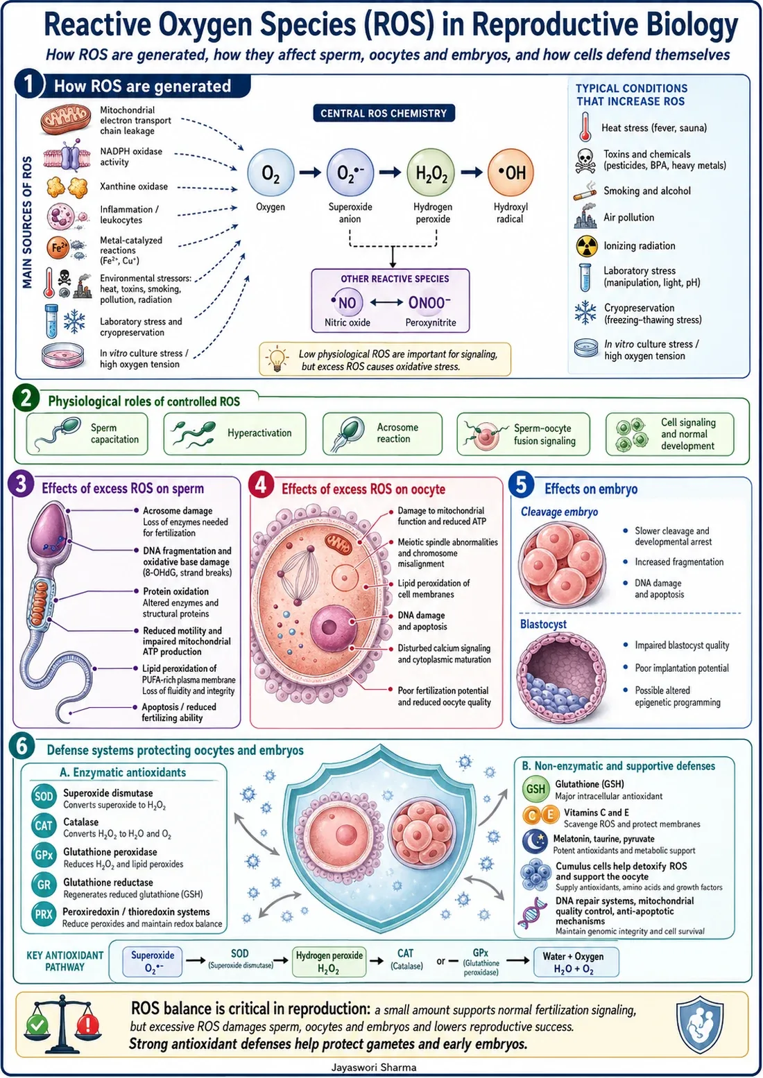
The goal is balance, not complete elimination. This is a point where I often disagree with how oxidative stress gets discussed, because ROS is usually presented purely as an enemy.
Controlled reactive oxygen species participate in sperm capacitation, hyperactivation, the acrosome reaction and normal cell signalling. A cell with no ROS at all would not function.
Excess is the problem, and excess has many sources: mitochondrial activity, inflammation, light exposure, high oxygen concentration, pollutants, prolonged handling, and unstable temperature or pH. Several of those are laboratory variables. That should get our attention.
The damage is specific to each cell type. In sperm, oxidative stress can damage membranes, motility and DNA. In oocytes, it can disturb mitochondria and the meiotic spindle. In embryos, it can slow cleavage, increase fragmentation and reduce blastocyst competence.
Cells do defend themselves. Glutathione, superoxide dismutase, catalase, glutathione peroxidase and peroxiredoxins form part of that defence system. But the defence has a capacity, and our job is to avoid exceeding it.
This is the section I would ask every new laboratory staff member to read twice. Stable gases, stable temperature, controlled pH, minimal light exposure and good air quality are biological protection. They are not laboratory decoration, and they are not optional refinements for well-funded centres only.
Key idea: A small amount of ROS supports signalling. Excess ROS damages the very cells we are trying to protect.
7. Why maternal age increases the risk of aneuploidy
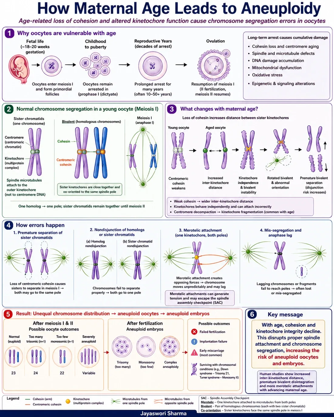
The oocyte may wait for decades before completing meiosis. When I first fully absorbed that, the age curve stopped being a statistic and became a mechanism.
Female germ cells enter meiosis before birth and remain arrested in prophase I until reproductive life. After ovulation, the oocyte pauses again at metaphase II until fertilization. No other cell in the body is asked to hold a partially completed division for thirty or forty years.
During that wait, cohesin proteins are responsible for keeping homologous chromosomes and sister chromatids correctly connected. With age, cohesion weakens and the geometry of sister kinetochores becomes less stable.
That instability increases the chance of premature separation, incorrect microtubule attachment, non-disjunction and lagging chromosomes. The result may be an oocyte or embryo with too many or too few chromosomes. Mitochondrial dysfunction, oxidative stress and altered signalling can add further pressure on a system that is already less reliable.
I want to state the clinical meaning carefully, because this is where patients are often failed by how we talk. Age-related aneuploidy is a structural and molecular problem inside the oocyte. It is not simply a number written on a patient file, and it is not a moral judgement about when someone chose to have children. Understanding the mechanism is what allows us to counsel honestly instead of vaguely.
Key idea: With age, weakening cohesion and less reliable kinetochore attachment make chromosome segregation more error-prone.
So why do we study cell biology again?
Because every visible IVF outcome is the final result of invisible cellular events.
A protocol tells us what to do. Cell biology explains why it must be done that way. Molecular biology helps explain why the expected result sometimes does not occur.
I have worked in environments where procedures were performed correctly by people who could not explain a single step of the reasoning behind them. It looks the same on a good day. It falls apart on a difficult one, because you cannot troubleshoot a process you do not understand. You can only repeat it and hope.
That is the real argument for going back to the organelles, the pathways and the signalling cascades. Not to pass an examination. To be the person in the laboratory who can say why something failed, and what to change.
A skilled embryologist does not only handle cells. A skilled embryologist understands what those cells are trying to achieve.
If you work in an IVF laboratory and want to discuss any of this, or if you are a student trying to connect your cell biology coursework to clinical practice, I am always happy to hear from you.